Western blot

Principal

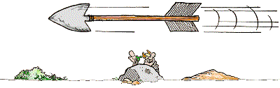
TRANSFERENCE OF PROTEINS ON A MEMBRANE AND SPECIFIC DETECTION WITH ANTIBODIES

The process can be divided into 3 steps:
1) Transfer of the protein on the membrane (blotting)
2) Saturation of free binding site of the membrane (blocking)
3) Specific detection of Your Favorite Protein (YFP) with antibodies

A nice animation was made by Lodish et al. ©:
Western blot, Lodish


1) Blotting

The proteins, separated by a SDS-PAGE, are transfered onto a membrane applying an electric field.

Transfer-membranes

The main characteristic of the membrane is that it adsorbs protein. Therefore, it is important not to touch those membranes with your fingers (use gloves!). Make sure that gel, membrane and paper are from the same size: large overhangs can prevent efficient current and transfer of the proteins. The membranes as well as the filterpaper are pre-soaked with Transfer-buffer (PVDF-membranes are additionally activated with MeOH previously) for ~ 15 min. Also the gel should be equilibrated for 15 min in ice-cold Transfer-buffer (otherwise shrinking will occure resulting in a distorted transfer-pattern). The membrane once placed onto the Gel should not be moved further (protein can already adsorb). It is important not having air bubbles between the membrane and the gel: roll out the membrane e.g. with a glass test tube. This also reduces the moisture between gel and membrane for having better contact between both and thus better transfer.


PVDF (PolyVinylidene DiFluoride):
PVDF-membranes have a very high protein binding capacity (170-200[600] µg/cm2), thus it is more prone for higher background.
To activate the PVDF-membrane soak it for 1-2/[15(?)] min in MeOH before use (do not let them dry afterwards or re-soak).
MeOH can be completely removed from the Transfer-buffer (e.g.for the transfer of large proteins) - but nevertheless, activate PVDF-membrane with MeOH.

Nitrocellulose (NC):
Be careful, NC-membranes are brittle when dry, thus supported NC-membranes are recommended.
NC-membranes have a high protein binding capacity (80-100[180] µg/cm2), non-specific protein binding sites are easily blocked (low background) and are much cheaper than PVDF-membranes.
With 100 µg protein per lane, each band has less than 10 µg, thus the protein binding capacity usually is more than enough.
To activate NC-membranes, place them *on* the surface of the Transfer-buffer, wait until they got grey, then submerge them into the buffer. The Transfer-buffer requires MeOH for better binding characteristics. For small proteins (< 15 kDa) 0.2 µm pore size is recommended, for big proteins (> 100 kDa) SDS can be added to the Transfer-buffer to increase the mobility (but it decreases the binding on NC).
In the CBL we use Whatman Protran NC-membranes with 0.45 µm pore size, precutted (7 x 8.5 cm), and prepackaged into a sandwich with 2 sheets of 3MM blotting paper; portein binding capacity is stated to be 80 µg/cm2 (BA85/3MM, #10485374).

Blotting system

Principally 3 types for blotting are possible: Wet, Semi-dry and Dry.
In any case a sandwich is formed from: cathode (-) - (sponge) - paper - gel - membrane - paper - (sponge) - anode (+).
The Wet or Tank blot has less harsh conditions and is util for large proteins (> 100 kDa), but requires bigger quantities of transfer buffer and is slower than the Semi-dry blot; on the other side, the Semi-dry blot generates more heat, which can damage the protein (detection) and colour the membrane. The Dry blot is not very common.
Generally, the transfer of large proteins (> 100 kDa) is slow, they tend to precipitate within the gel, so adding 0.1% SDS is recommended. MeOH tends to remove SDS from the proteins, thus a final MeOH conc. of 10% (instead of 20%) can be a good choice (or PVDF-membranes can be used w/o MeOH).
For small proteins the SDS can hinder the binding of the proteins on the membrane - thus it may good to remove the SDS completely from the Transfer-buffer and keep the MeOH at 20%.
Before the run, gel and membrane should be equilibrated for 15 min in ice-cold Transfer-buffer (otherwise shrinking will occure resulting in a distorted transfer-pattern). During the run the ice-cold buffer should be stirred (add a magnetic bar and place system onto a magnetic stirrer) to maintain uniform temperature and thus conductivity.

Wet/Tank:
In the CBL we have the Wet-blot "Mini Trans-Blot"-system (#170-3935) which is compatible with the SDS-PAGE Mini-Protean Tetra Cell (#165-8001) from Biorad.

Transfer-buffer:
Several recipies exits - indicating that the exact composition is not too important.
Only for Semi-dry systems the pH should be below 8.5 for not damaging the graphite electrodes.
The universal Transfer-buffer is: 25 mM Tris, 192 mM glycine, 10-20% MeOH, w/o SDS (use up to 0.1% SDS for proteins > 80 kDa). Do not adjust pH, should be ~8.3.

Blotting:

Prepare the gel sandwich
The blue cooling unit has to be froozen @ -20 °C
The Transfer-buffer has to be stored at 4 °C
Use gloves, do not touch the membrane
Gloves
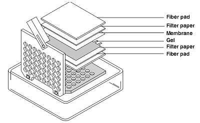
 1) Cut the filter paper and blotting membrane to the size of the gel (10 x 7.5 cm)

 2) Soak NC-membrane, filter paper and fiber pad for 15 min in ice-cold Transfer-buffer
   Equilibrate the gel for 15 min (not longer!) in ice-cold Transfer-buffer
   PVDF-membranes have to be activated with MeOH for 2(15?) min previously

 3) Place cassette with grey side down into a box
   Place soaked fiber pad on Cassette
   Place soaked filter paper on fiber pad
   use 1 filter paper from Biorad or 3 thin filter papers
   Place equilibrated gel on filter paper
   Place soaked membrane on gel
   Use glass tube to remove air bubbles
   Place soaked filter paper on membrane
   Place soaked fibre pad on filter paper
   Close clear tap and fix the gel sandwich
   Do not move sandwich during locking the Cassette

Gel Sandwich
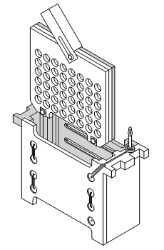
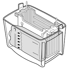
 4) Place the Cassette into the module:
   grey side of the Cassette to the black part of the Module
   (do the same for the second cassette, if necessary)

 5) Place the Module into the Tank
   Beware the orientation:
   Black side of the Module to the black mark from the Tank

 6) Place the frozen blue cooling unit in front of the Module into the Tank

 7) Fill into the Tank the Transfer-buffer up to the "Blotting"-mark (~ 450 ml?)

 8) Place a magnetic stirrer into the Tank. Keep buffer stirred during blotting.

 9) Connect system to Power supply (beware of the orientation / colour code)

10) Start blotting:
   12 hours: 30 V, constant 100 mA (36 Wh)
    3 hours: 60 V, constant 200 mA (36 Wh)
    1 hour: 100 V, constant 350 mA (35 Wh)

use constant current: if the ionic strength gets too high, the voltage drops,
thus preventing overheating; only the transfer time increases much.


(images are taken from the instruction manual)
Sandwich mounting Blot assembly

11) Stop the blotting after the indicated times and decompose the gel sandwich.

12 a) PVDF-membrane:
Let them dry completely (fixes proteins on membrane), then rehydrate with MeOH and Transfer-buffer. Afterwards place membrane with gel-faced side upwards onto TBS(T).
12 b) NC-membrane:
Place membrane with gel-faced side upwards onto TBS(T).

Optional: Make Coomassie staining for the gel to prove the complete emigration of the proteins out of the gel. This does not prove that all those proteins have adsorbed onto the membrane.


2) Blocking

As the membrane generally adsorbs proteins, hence it adsorbs also antibodies used for detecting YFP (background). Thus, before applying the antibody the non-specific protein-adsorption capacity has to be saturated by adding a not-interferring protein (lowering background). On the other side, too stringent blocking can release YFP from the membrane.
Usually cheap protein is used, like:
a) non-fat milk-powder: Cheap. Contains phospho-casein (can not be used for detecting phospho-proteins), biotin (not compatible with Streptavidin)
b) BSA, Cohn fraction V

10x TBST: 12.1 g Tris-HCl, 80.0 g NaCl, pH 7.6, 5 ml Tween 20 ad 1,000 ml
Use 3% BSA or 5% milk-powder in TBST (= blotto). Mix well and filtrate.
Incubate membrane 1 h @ RT.

Buffer
Stringency (5+)
Comments
TBST
+
cheap, w/o biotin; some background, may release prot. from membr.
TBST, 1% Tween or
  Triton X-100
++
cheap, w/o biotin; some background, may release prot. from membr.
Blotto
  5% milk powder in TBST
+++
cheap, w/o background; w/ biotin, may release prot. from membr.
1-3% BSA
  Cohn V grade in TBST
+++
w/o background; expensive, w/ biotin
Biotin inteferes with (strept)avidin detections system
N3- for stabilizing the buffer interferes with HRP detection systems
Pi based buffers (like PBS) interfere with AP detection systems

Blocking:

13) Prepare 50 ml(?) Blotto for each membrane.
14) Transfer 30 ml Blotto into a clean tray.
15) Place membrane with gel-faced side upwards(?) into Blotto.
16) Incubate 1 h @ RT, shaken (gyro-shaker)
Gloves

under construciton


Detecting

At the beginning antibodies should not be too diluted; the best conc. has to be tried out. As an orientation use:
Polyclonal serum or IgG-solutions: 1:100 - 1:1,000
polyclonal (affinity) or monoclonal Ab: 1 - 10 µg/ml
Required buffer volume: 250 µl/cm2 (10 x 8 = 80 cm2: 20 ml)
When membrane in sealed bag: 100 µl/cm2 (10 x 8 = 80 cm2: 8 ml)

Primary Antibody

Dilute primary antibody in TBST according manufactors instructions (usually 1:1,000 - 1:5,000). Too much antibody result in non-specific bands; this can be reduced by stringent blocking conditions but also blocking-protein can reduce sensitivity. With not enough antibody you may not detect YFP.
Polyclonal Ab can detect different epitopes. May be an advantage, when not proved to be suitable for WB.
Ab-buffer: TBST or blocking buffer. Incubation time: some hours to overnight, preferred @ 4 °C with agitation and antibiotic.
Afterwards, wash several times with TBST.

Primary antibody:
Mouse α-P-selectin = α-CD62P (Santa Cruz, #sc-18834): 200 µg/ml monoclonal IgG1. Storage: 4 °C
  WB: 1:1,000 (Predicted molecular weight: 91 kDa). Weight of P-selectin: 140 kDa
  IP: 1-2 µg/ml cell lysate
  IF: 1:50-500; 1 µg/106 cells

Mouse α-GST (Pierce, #MA4-004): 100 µg/ml monoclonal IgG1. Storage: -20 °C
  WB: 1:1,000 (Predicted molecular weight: 26 kDa). Weight of GST: 26 kDa
  IP: 1-2 µg/ml cell lysate

Secondary Antibody

Dilute secondary antibody in TBST according manufactors instructions (usually 1:1,000 - 1:20,000). Too much antibody result in non-specific bands; this can be reduced by stringent blocking conditions but also blocking-protein can reduce sensitivity.
Ab-buffer: TBST or blocking buffer. Incubation time: 1-2 hours, preferred @ RT with agitation.
Afterwards, wash several times with TBST.

Secondary antibody:
Goat α-Mouse-HRP (Millipore/Chemicon, #AB181P): polyclonal, affinity purified, lyophilized, Storage: 4 °C
Reconstitute: add ... µl steril A. dest., incubate 1-2 h @ RT. Centrifuge.
Long term storage: aliquot, add 50% steril glycerol and store @ -20 °C

  WB: 1:5,000-100,000

Conjugation

Several reactions exist to detect the secondary Ab:
POD or HRP (Horseradish Peroxidase):
Chromogene
Sensitivity (6+)
Comments
4-chloronaphtol (4-CN)
  + H2O2
+
cheap, blue/black bands; intensity diminish fast
3,3'-diaminobenzidine (DAB)
  + CoCl2 + H2O2
+++
cheap, black bands; high background
Chemiluminescence
  ECL or (ECL+)
++++
(+++++)
very sensitive, good documentation; expensive, requires system for develop fotos
DAB: Detection limit: 5 ng of protein. Suggested dilutions: 1° Ab 1:500, 2° Ab 1:2,000-20,000
Stock: 1% DAB (20x) in A. dest.: 100 mg DAB in 10 ml A.dest. Add ~5 drops of 5 N HCl to turn colour brown. Shake for 10 min. Aliquot in 1 ml and store at -20 °C
0.3% H2O2 (20x): 100 µl 30% H2O2 in 10 ml A. dest. Working soln: 0.05% DAB (10 drops) + 0.015% H2O2 (10 drops) in (10 ml) 0.01 M PBS (pH 7.2): beware pH - pH < 7.0 low staining, pH > 7.6 cause background staining.
Incubate membrane @ RT for 10-15 min

AP (Alkaline Phosphatase):
Chromogene
Sensitivity (6+)
Comments
5-bromo-4-chloro-3-indolyl phosphate (BCIP)
  + Nitro blue tetrazolium (NBT)
+++
cheap, good documentation; requires DMF as solvent, intensity diminish slowly

Generally: Secondary antibody should be highly cross-absorbed to serum proteins and affinity purified.
Lyophilized antibody should be reconstituted to approx. 1 mg/ml.
HRP: 40 kDa, pH 5-7, fast reaction, good stability; inhibited by N3-, CN-
AP: 140 kDa, pH 8-10, inhibited by Pi, CN-, EDTA.

In the CBL we use HRP with DAB (
Pierce #34002).
25 ml DAB Stock solution (10x); 250 ml HRP Substrate buffer. Storage: 4 °C.
Immediately before use: Add to 22.5 ml HRP Substrate buffer, 2.5 ml DAB Stock solution, mix thoroughly.

Detection

17) Wash 3 x 10 min @ RT with 50 ml TBST.
18) Primary antibody: dilute in ~20 ml (8 ml in a bag) TBST (or Blotto) and incubate 1-6 h @ RT, shaken (gyro-shaker).
19) Wash 3 x 10 min @ RT with 50 ml TBST.
20) Secondary antibody: dilute in ~20 ml (8 ml in a bag) TBST (or Blotto) and incubate 1-2 h @ RT, shaken (gyro-shaker).
21) Wash 3 x 10 min @ RT with 50 ml TBST.
22) Add activated DAB solution to membrane and incubate @ RT until desired development is archieved (typically 5-15 min)
Gloves


under construciton


Troubleshooting

No signal
Poor protein-transfer onto the membrane: make sure
  - sufficient contact between gel and membrane (not too much buffer)
  - the correct activation of the membrane and the correct assembly of the sandwich
  - not too short (counterstain gel w/ Coomassie) or long (observe Rainbow size marker) transfer time
  - that
Insuficient binding to membrane:
  - MeOH is required for binding to NC-membrane, but at high conc. it removes SDS, especially from big proteins
  - small proteins may break through: use smaller pore size
Not enough primary or secondary antibody bound: use more antibody and longer incubation time.
Secondary antibody does not recognize primary antibody.
YFP has not lost antigenicity during process. Use positive control.
Insuficient antigen:
  - use at least 20-30 µg protein per lane.
  - too much washing of the membrane.
  - blocking-buffer has masked antigen.
Use of N3- which is incompatible with HRP
Old, inactive substrate for detection

Uniform high background
Insuficient blocking: use 5% non-fat dry milk or 3% BSA in TBST for at least 30 min. Use this also as antibody buffer.
Too high antibody concentration, too high incubation temp., or too long incubated.
Inspecific reaction of (primary/secondary) antibody with blocking reagent: test by using only 2° antibody
Cross reaction between blocking reagent and (primary/secondary) antibody: use other blocking protein.
Insufficient washing of antibody: increase washing cycles, use Tween 20.
Membrane problems: NC has less background than PVDF. Make sure that membrane was activated previously.

Speckled background
Antibody added directly onto the membrane.
Aggregate formation in HRP conjugate: filter conjugate through 0.2 µm filter.
Contaminated equipment: make sure that all equipment and trays are clean.
Membrane problems: use gloves and forceps.

Multiple bands
Cell lines frequently passaged: gradually different proteins acculumate.
Multiple forms of YFP: phosphorylated, methylated, acetylated, ubiquitinylated, glycosylated.
YFP has been degraded => use protease inbhibitor.
(unreported) splicing variant.
Too high (primary or secondary) Ab.
Usage of non-purified Ab.

Others
White spots: Air bubbles.
Black spots: Ab reacts with blocking protein.


under construciton

back


Last modified: 21.03.2012