Cytotoxicity


Studying cytotoxic effects means determining harmful effects on cells (in-vitro).
The aim can be:
- to prove the intoxicity (safety) of pharmaceutical or cosmetical products or food-stuff
- to screen for new biological active compounds
- to prove the biocompatibility of (medical) materials

The advantages of using cell culture are:
- use of human cells (and not from animals)
- reduce the number of animal 'consuming' experiments
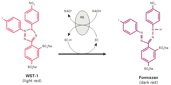
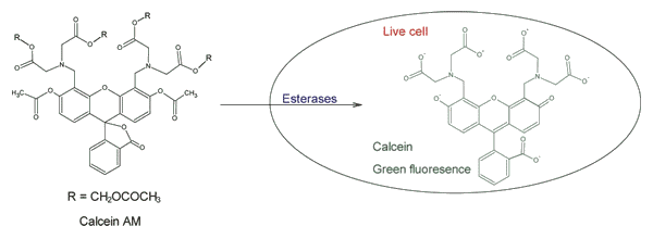
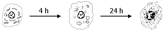
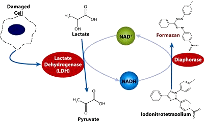
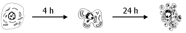
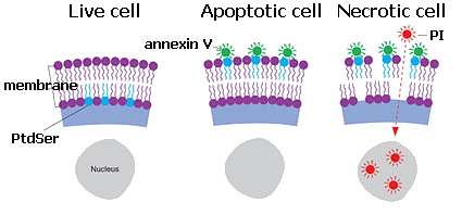
- cellular effects can be detected
- automatization is possible (high throughput screening)

as well the disadvantages are:
- systemic effects are not (or only difficult) detectable
- no long-term studies
- metabolization of components can only be simulated
- a correlation between the in-vitro and in-vivo results is required

Finally, experiments with animals can not be substituted but at least reduced.

Mouse-culture
This is still not possible.

Recommendations

- Use cell lines only from authorized collections, e.g. ATCC, HPA or DMSZ
- do not subcultivate more than 5 times from the reference line, in order to avoid genetic drift
- do not use antibiotics routinely

Consider also:
- use of mortal (primary) vs. immortal (cancerigenic, but reproducible) lines.
- defined media (which additives are necessary?) vs. complete/complex media (using FBS, not reproducible).
- for the probing substance: which concentration, which solvent, syn- or antagonistic effects?
- presence of FBS during contact (may adsorb probing substance).
- the time period of contact (e.g. 1, 5, 24 h) and medium change afterwards.
- time point of analysing the effect (e.g. after 6, 24, 72 h).
- effect should be analysed by 2 independend methods.
- the used cell line is representative for the organ to study?

Control:
- Blank: Medium w/o cells
- Neg. control: Medium w/ cells but w/o sample (growth control).
- Mock control: Medium w/ cells and w/ solvent for test substance.
- Pos. control: Medium w/ cells and with known effect-causing substance.

Cell lines

Usefull cell lines, representative for the function of the respective organ are:
Skin (simulating contact): HaCaT
Intestine (simulating absorption): Caco-2
Liver (simulating detoxification with P450-system): THLE-3
Kidney (simulation detox/reabsorption of proximal nefron): HK-2

Other cell lines:
Breast cancer: MCF-7
Immune system (monocytes): THP-1


1) Test for viability/survival

With these tests metabolic active cells are detected. Typical tests are:
1.1) Tetrazolium: detects dehydrogenase activity.
1.2) Sulforhodamine B (SRB): detects protein biosynthesis.
1.3) C12-Resazurin (Alamar blue): detects dehydrogenase activity.
1.4) Calcein-AM: detects esterase activity.
Methods overviewpaper

Positive Control:
H2O2 for wound disinfection (35 g/l = 1 M)
Add 50 µl/ml = 50 mM.

Take into account the general considerations and adapt the protocol accordingly!

1.1 Tetrazolium

Different tetrazolium salts (INT, MTT, XTT, MTS, WST) can be reduced by formed NADH from metabolic active cells, resulting in a different coloured formazan, which can be detected by a photometer. As most of the NADH is formed in the mitochondria, 1-methoxy-phenazine methosulfate is needed as an electron-carrier to transfer the reduction equivalents from the mitochondial matrix into the medium. Under normal conditions the formed colour correlates linear with the cell number (use for cell count) or under different conditions the (less) formed colour indicates cell inhibition or death (use for cytotoxicity).

Consider:
Stressed cells (e.g. by Actinomycin D 10 ng/ml for 20 h) can also exhibit a higher metabolic activity than normal but they are not proliferating, thus the effect would be more cytostatic and not cytotoxic.
Each formazan has its charactertistics.
- INT: Abs. 490-520
- MTT: Abs. 550-600 nm (565 nm), 4 h incubation, insoluble formazan (requires solubilisation step).
       Phenol red (from medium) may interfere with absorbance.
       It is also reduced by the GSH-transferase thus it is not adequate, when sample interfere with GSH metabolism.
- XTT: Abs. 450-500 nm (460 nm), 2-4 h incubation
- MTS: Abs. 495 nm
- WST-1: Abs. 420-480 nm (438 nm), 0.5-2 h incubation, stable, water soluble formazan
Measure background against 690 nm.
WST reaction
For convenience we use WST-1 (Clontech #630118).

Protocol:
First use:
Thawed WST-1 ready to use solution at 37 °C, mix very well to dissolve precipitates, aliquot to 1.2 ml in Eppendorf-tube and store at -20 °C, dark.

1) Add 1 ml of medium (e.g. DMEM, 5-10% FBS, 10 mM HEPES) into each well of a 12-well MTP
2) Inoculate with 4 * 104 cells (~ 40 µl) and incubate 24 h (cell attachment).
3) Add test substance at different concentrations. Incubate for 24 h (or desired time).
4) Thaw up the WST-1 aliquots at RT in the dark, mix very well to dissolve precipitates.
   If test substance reacts with WST-1, remove medium, wash cells 1-2x with PBS and add 1 ml fresh medium
5) Add 15-25 µl WST-1 and incubate cells for 0.5-4 h (dark).
6) Calibrate photometer against medium (blank) at λ 440 nm and λ 690 nm.
7) Measure Abs. at λ 440 nm and λ 690 nm for each sample.

For 96-well MTP and 100 µl medium:
4) Thaw up the WST-1 aliquots at RT in the dark, mix very well to dissolve precipitates.
   Mix 200 ml WST-1 with 10 ml fresh medium.
5) Remove medium (wash cells 1-2x with PBS if necessary) and add 100 µl medium containing WST-1.
6) Incubate cells for 0.5-4 h (dark).
7) Measure absorbance at λ 440 nm and λ 690 nm for each sample using an ELISA-reader.

1.2 Sulforhodamine B (SRB)

SRB binds under mild acidic conditions to basic amino acids, thus it can be used to detect protein biosynthesis. At neutral pH SRB desorbs again, which makes it accessible for its determination. In the CBL until now we have not introduced this method.
SRBpaper

Principally:
- fix cells with TCA (10% final).
- incubate 1 h @ 4 °C.
- wash with water (to remove TCA).
- add SRB (0,4%).
- incubate 30 min @ RT.
- wash with 1% HAc to remove excess SRB.
   Do not wash too much in order to avoid desportion of dye from protein.
- neutralize with 10 mM Tris (pH 10.5) to desorb and solubilize protein bound SRB.
- incubate (shaking) 5 min @ RT.
- determine absorbance @ 560 nm. Substract background measured at 690 nm.
Sulforhodamine B

1.3 C12-Resazurin (Alamar blue)

C12-Resazurin is a redox indicator, which can be reduced to Resorufin by NADH, means by metabolic active cells (dehydrogenase activity). Resorufin exhibits fluorescence (563/587 nm) and can be detected by epifluorescence microscopy or photometrically.
Troubleshooting: Cys, DTT and tiols reduce resorufin to the colourless dihydro-resorufin.

Principally:
- add C12-Resazurin to the culture medium (5 µM final).
- incubate 15 min (up to 4 h?).
- wash with PBS.
- determine absorbance @ 570 nm or fluorescence (563/587 nm).
(Substract background measured at 690 nm?)
Resazurin

1.4 Calcein-AM

Lipophilic Calcein-AM permeates across the membrane into the cells, where it is hydrolysed by esterases to green fluorescence Calcein (470/520). Thus this method detects metabolic active cells (esterase activity). Calcein, which get liberated into the medium, can be quenched by 5 µM Co2+.

Principally:
- wash cells with PBS.
- resuspend cells in special medium.
- add Calcein-AM to final conc. of 1 µM.
- incubate 30 min @ 37 °C.
- determine absorbance @ ... nm or fluorescence (470/520 nm).
(Substract background measured at 690 nm?)
Caclein-AM


2) Test for (cellular) necrosis

With these tests necrotic cells are detected, means cells who suffered a death by "catasrophe". Typical characteristics for a beginning necrosis (0,5-4 h) are swelling of the cells, followed by a disruption of the cell membrane (cells become leaky). Then the metabolism stops, the organelles are not functional, resulting in an energy failture, which terminates in cell lysis (24 h). This release of cell components usually causes an inflammatory reaction.
Cytotox assayspaper           Compare assayspaper

necrosis
Tests for necrosis usually detect the liberation of cytosolic content into the medium. It is important to differentiate between (primary) necrosis and secondary necrosis, i.e. liberation of cell components into the medium out of apoptotic bodies, which are not cleared in cell culture as they are in-vivo. Typical tests are:
2.1) Trypan blue: detects membrane disintegration.
2.2) Neutral red uptake (NRU): detects membrane disintegration.
2.3) Propidium iodide (PI): detects membrane disintegration.
2.4) LDH-release: detects liberation of Lactate-dehydrogenase (membrane disintegration).

Positive Control:
H2O2 for wound disinfection (35 g/l = 1 M)
Add 50 µl/ml = 50 mM.

ROS inducer:
CuSO4
Stock: 10 mM CuSO4 * 5 H2O (Mw = 250 g/mol)
125 mg/50 ml, steril-filtrated or autoclaved.
Add 5 µl/ml = 50 µM and 25 µl/ml = 250 µM

Take into account the general considerations and adapt the protocol accordingly!

2.1 Trypan blue

Trypan blue (Biowest/Mayimex, #L0990) does not permeate across functional membranes into the cells, thus living cells are not stained while necrotic cells (with compromised cell membrane) are stained blue.

Protocol:
- resuspend (detached) cells in 100 µl PBS.
- add 10 µl trypan blue (0.5%) to 90 µl cell suspension.
- incubate 3-5 min @ RT.
- count cells microscopically (Neubauer).

Troubleshooting:
- do not stain longer than 5 min as trypan blue is toxic to cells.
- apoptotic cells are not stained.
- trypan blue exhibits a strong red autofluorescence.
Trypan bluepaper
Trypan blue

2.2 Neutral Red Uptake (NRU)

The test for NRU has the opposite principal as trypan blue: This permeates across functional membranes hence enters (among others) into the lysosomes, where it get protonated and changes its colour to red. Protonated neutral red can not leave the organlles again thus it accumulates. Living and early apoptotic cells are stained red.
We use Neutral Red Solution from Santa Cruz (sc-281691) or Sigma-Aldrich (#N2889) which has a concentration of 3.3 mg/ml = 0.33% in DPBS.

Principally:
Qualitative:
- add 50 µg/ml neutral red to the medium (15 µl/ml of 0.33% soln).
- incubate 2-4 h @ 37 °C (concentration and incubation time depends on the metabolic activity of the cells).
- wash cells 1x with PBS.
- count cells microscopically (Neubauer)

Quantitative:
- add 100 µg/ml neutral red to the medium (30 µl/ml of 0.33% soln).
- incubate 2-4 h @ 37 °C (concentration and incubation time depends on the metabolic activity of the cells).
- wash cells 2x with PBS
- extract neutral red with 800 µl 1% HAc in 50% EtOH (15 min)
- centrifuge supernatant: 3 min @ 10,000 g
- determine absorbance @ 540 nm. Substract background measured at 690 nm.
Neutral red

2.3 Propidium Iodide (PI)

PI has similar characteristics as trypan blue: it does not permeate across functional membranes into the cells, hence living or apoptotic cells are not stained while it enters into necrotic cells (with compromised cell membrane) where it intercalates with the DNA exhibiting a red fluorescence (535/617). This method is frequently used to counter stain necrotic cells to differentiate them from apoptotic (and living) cells.

Principally:
- add 100 µg/ml PI (1.5 µM) onto a cover slip.
- incubate 5-15 min @ RT (dark).
- wash with PBS.
- observe with epifluorescence microscope (535/617 nm).

Troubleshooting:
- RNA may interfere: can be degradated by treatment with RNase A.
Propidium Iodide

2.4 LDH-release

This test has the same objective as the tests before but a different approach: Through compromised cell membranes (necrotic cells) cytosolic content is released into the medium, where it can be detected. Usually cytosolic Lactate-dehydrogenase activity is determined, although some tests prefer Glc-6-Pi-DH. The advantage of LDH over Glc-6-Pi-DH is its higher stability in the medium, but on the other hand FBS (usually part of the medium) contains traces of LDH thus the FBS has to be heat-inactivated before use.
For convenience we use the LDH-test from (Clontech #630117).

Protocol (adapted to 12-well MTP and normal Photometer):
First use:
Catalyst bottle: Add 1 ml A. dest. and mix thoroughly (10 min); store @ 4 °C, dark (several weeks).
Dye solution bottle: thaw up, ready to use.

1) Reaction solution: Prepare just before use. Mix 4 µl Catalyst and 196 µl Dye soln
    for 12 reactions (á 1 ml): 50 µl Catalyst and 2250 µl Dye
2) Shake plate where cells are growing to distribute LDH within the medium.
3) Transfer 800 µl medium into Eppendorf tube.
4) Add 190 µl Reaction soln (contains INT, Lac, NAD+, diaphorase) and mix.
5) Incubate 30-60 min @ RT (dark).
6) Centrifuge Eppendorf tube (1 min, 8000 g).
7) Calibrate photometer against medium (blank) at @ 490 nm and @ 690 nm.
8) Take off 800 µl supernatant (w/o moving a possible pellet) and
9) Measure Abs. @ 490 nm and @ 690 nm for each sample.

Protocol (for 96-well MTP and ELISA-reader):
1) Reaction solution: Prepare just before use.
    for 96 reactions (á 100 µl): 200 µl Catalyst and 9 ml Dye
2) Shake plate where cells are growing to distribute LDH within the medium.
3) Transfer 90 µl supernatant into a new 96-well MTP.
4) Add 90 µl Reaction soln and mix.
5) Incubate 30-60 min @ RT (dark).
6) Measure Abs. @ 490 nm and @ 690 nm for each sample.
LDH release


3) Test for apoptosis

With these tests apoptic cells are detected, cells who suffered a programmed cell death. Typically, at the beginning (0,5-4 h) the cells are shrinking and forming blebbs (the organelles are within these blebbs), the nucleus starts to fragment but the cell membrane remains functional (means the energy metabolism does not stop and the cells do not become leaky). Later on (24 h) apoptotic bodies (containing functional organelles) are formed, the membrane remains intact thus cell components are not released into the medium; contrary macrophages and neighbour cells are phagocyting the apoptotic bodies preventing a release of cell components and thus preventing inflammatory reaction. In cell culture this very last step will not occure (or you are performing co-cultivation) thus finally the membranes of the apoptotic bodies get compromised and cell content is released (aka secondary necrosis).
Apoptosis Assays Review paper           Apoptosis Assay-pitfalls paper

apoptosis
The tests can differentiate three phases of apoptosis:
Early Apoptotic Phase:
3.1) Annexin V: PtdSer changes from the internal to the outer leaflet, where it can bind to annexin V
3.2) Cytochrome c: Cyt. c is liberated from the mitochonrion to the cytosol
3.3) Mitochondrial Membrane Potential (MMP)
3.4) Initiator Caspases: Caspase 8 (activated by external TNF-α, Fas), caspase 9 (activated internally by cytosolic Cyt. c)
Mid Apoptotic Phase:
3.5) Effector Caspases: Caspases 3, 6, 7, which are activated by the initiator caspases
3.6) Nuclear condensation
End Apoptotic Phase:
3.7) DNA-ladder: Fragmentation of DNA into ~200 bp fragments
3.8) Apoptotic bodies: The formation of apoptotic bodies can be observed by microscope

Additionally some links for commercial products can be found at apoptosisworld.com; protocols from researchers for researchers are available at molecularstation.com and protocol-online.org.

Positive control:
Camptothecin (BioVision #1039-1, 1 ml, 2 mM), 100 µl aliquot, store at -20 °C.
Add 1 µl/ml = 2 µM (should be enough, but it is not always like this...)
instead we use now 5 µl/ml = 10 µM.
Doxorubicin*HCl (Mw = 580 g/mol): 1.16 mg/ml = 2 mM, 100 µl aliquot, store at -20 °C.

3.1 Annexin V

Usually phosphatidyl serine is found only in the inner leaflet of the cell membrane, but at the very beginning of the apoptosis PtdSer is transfered to the outer leaflet. There, it can bind the protein annexin V in a Ca2+-dependent manner.
This can be detected by a) adding fluorescently marked annexin V (e.g. annexin V-FITC) or b) adding annexin V and fluorescently labelled α-annexin-antibody.

For convenience we use the annexin V test from Santa Cruz (#sc-4252 AK: has 5 ml 10x Assay buffer, 50 µg/250 µl Annexin V-FITC, 2 ml of 50 µg/ml propidium iodide, PI; enough for 100 tests).

Protocol:
Before use (for each cover glass):
Dilute 50 µl 10x Assay-buffer with 450 µl A. dest. (1x Assay-buffer)
Add to 100 µl 1x Assay-buffer 2.0 µl Annexin V-FITC and 10 µl PI
Prepare 1x PBS

 1) Place round cover glass into the required wells of a 12-well MTP.
 2) Add 1 ml of medium (e.g. DMEM, 5-10% FBS, 10 mM HEPES) into each well.
 3) Inoculate with 4 * 104 cells (~ 40 µl) and incubate 24 h.
 4) Add test substance at different concentrations. Incubate for 24 h.
 5) Take off medium and rinse cells with PBS.
 6) Wash cells once mit 400 µl 1x Assay-buffer.
 7) Place 100 µl 1x Assay-buffer (w/ Annexin V-FITC and PI) onto a piece of Parafilm
    and place cover slide up-side-down onto it.
 8) Incubate 15 min @ RT in the dark.
 9) Place cover glass on slide and observe fluorescence:
   - green filter (GFP/FITC) for annexin V-FITC (495/521).
   - red filter (rhodamine) for PI (535/617).
annexin V

Troubleshooting:
green: apoptotic cells; green + red: necrotic cells; w/o staining: healthy cells
measurement depends on Ca2+-conc.: usually not more than 1.8 mM; add only 10 min before analysis
- better wash with HEPES (10 mM HEPES/NaOH, pH 7.4, 140-150 mM NaCl, 5 mM KCl, 1 mM MgCl2, 1.8-2.5 mM CaCl2; sterile filtrated) than with PBS
- incubate on ice
- medium (DMEM, M199 is better than RPMI1640)

3.4/5 Caspase activation


Test for autophagy


Autophagy methodspaper


back


Last modified: 17.01.2014